Our broad interest is to understand how cell-cell signaling molecules set up spatial pattern, particularly in the development and regeneration of neuronal connections. We use a broad range of approaches including biochemical, molecular, cellular, and in vivo studies.
Wiring up the nervous system: molecular cues for axon guidance and regeneration.
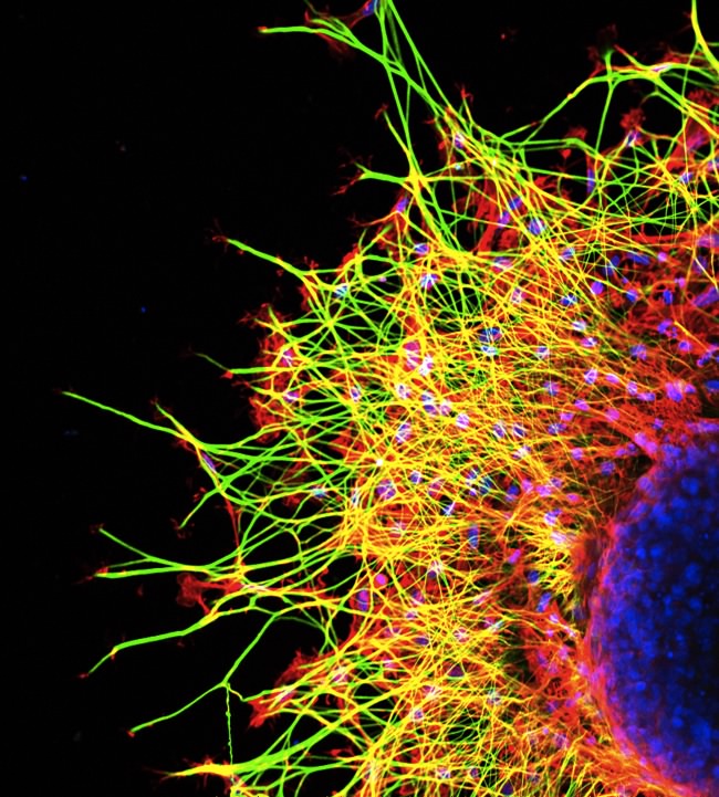 The functioning of the nervous system depends on the precise and complex spatial order of its connections. These connections are initially set up during development, when the motile growth cone at the tip of the axon is directed toward the correct target by axon guidance cues. Using methods developed in the lab [1], we have identified a number of novel extracellular cues or their receptors, and this can open the door to new research areas. We identified some of the first developmental axon guidance cues, members of the ephrin family, and showed that they provide gradients of positional information that guide the formation of neural topographic maps [2]. In addition to development, we are interested in axon regeneration. When connections in the adult central nervous system are lost due to injury or disease, regeneration is minimal, creating a major clinical challenge. Our work recently identified a receptor for the chondroitin sulfate proteoglycans, a major class of regeneration inhibitors in the central nervous system. This receptor-ligand interaction now provides a therapeutic target to promote CNS regeneration [3,5].
How is information from outside the cell converted to a spatially appropriate intracellular response? RNA-based mechanisms.
How do extracellular cues produce a spatially appropriate response within the cell? We are particularly interested in RNA-based regulatory mechanisms. Regulation of mRNA translation provides a way to target protein synthesis to specific locations within the cell. 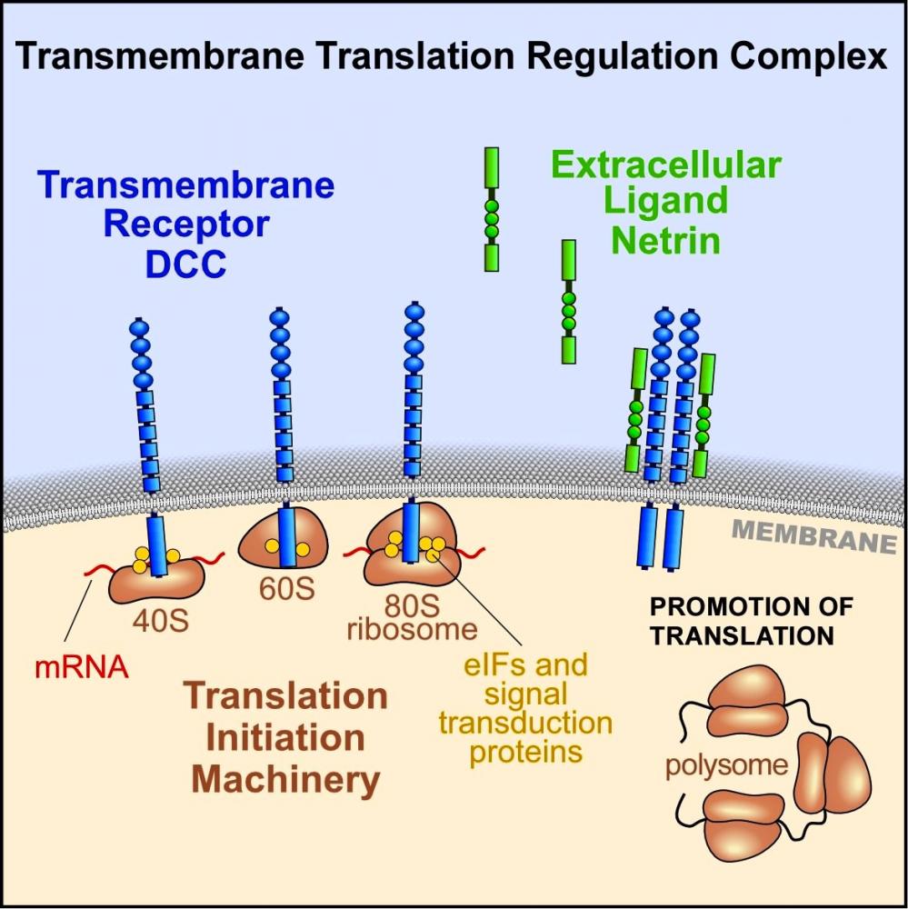 Since the neuron is a highly polarized cell, it provides an excellent model system to study localized translation. We and others have shown that protein synthesis can be regulated within the neuron in a precisely localized manner, and that it plays key functional roles in axon guidance and synapse plasticity. In recent work, we identified a generalizable mechanism where transmembrane receptors associate with protein synthesis machinery, providing a way for cells to localize and regulate translation in response to extracellular cues, based on a transmembrane translation regulation complex [4]. We are continuing to study RNA-based mechanisms by which extracellular cues control cell behavior, as well as functional roles for these mechanisms in axon guidance and cell polarity.
References
[1] Flanagan, JG, Cheng, HJ (2000) Meth. Enzymol. 327: 198-210 [review].
[2] Flanagan, JG (2006) Curr. Op. Neurobiol. 16:59-66 [review].
[3] Shen et al. (2009) Science 326:592-6.
[4] Tcherkezian et al. (2010) Cell 141:632-44.
[5] Coles et al. (2011) Science 332:484-488.